Ignoring This Canine Eye Condition Could Lead to Blindness
As alarming as it is to first notice this condition in your pet's eyes, not acting swiftly could be a big mistake. Find out why cherry eye, or prolapse of the lacrimal gland inside the third eyelid, requires prompt professional attention for the sake of your pet's eyesight.

Reviewed by Dr. Becker


Join the Bark & Whiskers™ Family
Sign up today for our FREE newsletter, packed with expert advice and insider tips to keep your beloved pet in tip-top shape.
View our Privacy Policy and Terms of Service.


STORY AT-A-GLANCE
- Dogs have a third eyelid that sometimes develops a condition called cherry eye, which is the descriptive term for prolapse of the lacrimal (tear) gland inside the third eyelid
- This third eyelid, or nictitating membrane, is designed to produce tears, keep the cornea clean and protected, and produce antibodies to fight infection
- Your dog’s third eyelid shouldn’t be visible; if it becomes visible, it signals a problem such as cherry eye that requires prompt examination by your veterinarian
- Cherry eye can sometimes be medically managed, but often requires surgical correction (but never removal of the lacrimal gland)
Editor's Note: This article is a reprint. It was originally published February 01, 2021.
Many a dog parent has had the unsettling experience of suddenly noticing a red bump or protrusion from the corner of their pet's eye. The common name for this condition is "cherry eye," which is a more descriptive term than the scientific "prolapsed gland of the nictitans." Nictitans means nictitating membrane, which in dogs is actually a third eyelid.
In case you weren't aware, dogs have not two, but three eyelids. There's the upper lid, the lower lid, and the third eyelid aka the nictitating membrane aka the haw. Third eyelids are essentially the same in different breeds and sizes of dogs, although the pigmentation can vary from breed to breed. Also, some are clear while others are cloudy.
Other types of animals also have nictitating membranes, including cats, birds, reptiles, fish and camels. The third eyelid helps keep the eyes moist in the presence of wind, sand or dirt — no blinking necessary. Needless to say, this can come in very handy when hunting for prey that can disappear in the blink of an eye.
Your Dog's Third Eyelid Has Several Functions
Your dog's third eyelid is a thin, opaque tissue that sits in the inner corner of each eye, below the lower lid, and serves four important functions:1
- It acts as a windshield-washer for the cornea (the clear surface at the front of the eye), keeping it free of debris and mucus
- It contains a lacrimal gland that produces about one-third of your dog's tears
- It contains lymphoid tissue that produces antibodies to fight infection
- It protects the cornea from injury
Interestingly, human eyes function in a similar way, but with two eyelids instead of three. When your dog's third eyelid closes, it can appear as though his eye is rolling back in his head. And sometimes when dogs sleep, their upper and lower eyelids open, revealing the closed third eyelid or "white eyeball" as some startled pet parents describe it!
In most cases, the third eyelid remains retracted. If it becomes visible, it can be a sign of a problem that requires prompt investigation by a veterinarian. For example, if you suddenly notice your dog's third eyelid for the first time, it could mean his eyeball has sunken into its socket due to severe eye pain.
It could also mean there's been an injury to the eye, or that the structure that holds the third eyelid in place is damaged or weakened. Other causes of a visible third eyelid include allergic conjunctivitis and autoimmune disease.2
In some dogs, a portion of the third eyelid is always visible — a condition called haws. When a dog is born with the third eyelid visible, this is a normal condition and does not indicate illness (although it is considered undesirable in show dogs).3
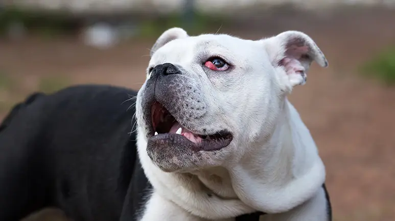
Cherry Eye
The lacrimal gland inside the third eyelid is held in place by tiny tissue fibers. Prolapse can occur in one eye or both, and once one eye is affected, there's a very good chance the other eye will be as well.
When a prolapse happens, you'll see red, thickened, irritated tissue in the corner of your dog's eye. Once it prolapses, the gland can grow increasingly inflamed and even infected. Your dog may seem to be managing just fine, and fortunately, the condition isn't typically painful. However, because the gland is no longer seated in its normal position, it can prevent adequate lubrication of the eye, which can lead to dry eye.
Any dog of any age can develop cherry eye, but certain breeds are more prone to the condition than others, including many of the brachycephalic (flat-faced) breeds, as well as the Beagle, Bloodhound, and Bull Terrier. Other breeds with the tendency include the Cocker Spaniel, Saint Bernard, and Shar-Pei.
The cause of third eyelid gland prolapse isn't well understood, but it's believed to be related to a connective tissue weakness in the ligaments that hold the gland in place.
Treatment Options
A prolapsed lacrimal gland can be treated either medically or surgically, however, it's crucial that a medical approach takes place quickly and aggressively. Treatment should begin as soon as the prolapse occurs and definitely within the first couple of weeks. In cases where a gland has been prolapsed for an extended period of time, there's usually no hope for nonsurgical intervention.
My approach with a dog who has just developed cherry eye is to immediately institute an aggressive protocol of topical compresses (steep an organic green tea bag for 30 seconds, then put in the refrigerator until cold, gently apply over the eyelid for 3-5 minutes, repeat with a fresh teabag 3 times a day), eye drops that contain N-acetyl carnosine, specific homeopathics, and nutraceuticals (including organic collagen and MSM) to control the inflammation and try to reestablish the integrity of the ligaments that hold the gland in place.
If medical treatment fails or isn't a viable option, a surgical procedure is required to seat the gland back in its normal position under the lower eyelid. One technique, called pocket imbrication, involves creating a new pocket in the tissue of the third eyelid to house the gland. Since there will be inflammation after the procedure that will take time to completely resolve, surgical results won't become fully evident for several weeks.
Another technique, called an orbital rim tack, involves suturing the gland to the tissues surrounding the orbital bone.
Unfortunately, these surgeries occasionally can and do fail, and a second or third procedure is necessary. How well things progress after surgery depends on the chronic nature of the condition and/or how inflamed the eye tissue is. Sometimes, lubricating eye drops are needed to supplement the tear film seasonally or periodically, throughout the rest of the dog's life.
Surgery to remove the lacrimal gland was once commonplace, but thankfully is no longer considered an acceptable form of treatment because it removes about half the lubrication capacity of the eye. Removing the gland is easy, but not a viable solution, long-term, without compromising the integrity of the eye. Removing the gland very often leads to keratoconjunctivitis sicca (KCS), or dry eye.
To prevent problems in the future, up to and including blindness, dogs with KCS must depend on their owners to manually lubricate their eyes for the rest of their lives. If your veterinarian suggests removing the gland as a form of "treatment," get a second opinion with a more up-to-date, qualified surgeon.
There's really nothing you can do to prevent cherry eye, but if your dog develops the condition, it's important to make an immediate appointment with your veterinarian to begin medical treatment, hopefully preventing the need for surgery, and restoring the health of your pet's eyes.
Sources and References
- Texas A&M Veterinary Medicine & Biomedical Sciences, December 3, 2020
- 1,2 Animal Planet
- 3 PetMeds